

A great deal of progress has been made in terms of materials, techniques and design of dental implants since the beginnings of modern implantology over 50 years ago. While titanium and titanium alloys have always been in use, the search for metal-free implantable materials began in the late 1960s and early 1970s, and during the last decade, zirconia has emerged as the most reliable implantable bioceramic. The International Academy of Ceramic Implantology (IAOCI) is an organization entirely dedicated to ceramic and metal-free alternatives to metal implants. It was founded in 2011 by Dr. Sammy Noumbissi, with whom Dental Tribune Online had the opportunity to speak about the mission and vision of the IAOCI, as well as the state of ceramic implantology today.
Dental Tribune Online: Dr. Noumbissi, could you please provide some background information on the development of ceramic implants?
Dr. Sammy Noumbissi: The use of dental implants to replace teeth has increased very rapidly in the last 15 or more years. With this increase in dental implant procedures, the number of manufacturers has increased too. Also, we have witnessed the introduction of various alloys of titanium over time.
Now, just like with any pharmaceutical or medical product, the increase in demand and changes in production methods come with problems and challenges. Although initially anecdotal, reports of titanium and titanium alloy intolerance have increased and are increasingly being investigated and demonstrated in the scientific dental literature. Based on the body of research available today, this intolerance of implant alloys can in great part be attributed to the release of metal ions in the host bone and surrounding tissue as a result of the breakdown and corrosion of metal alloys in the presence of body fluids and the oral environment in particular. Such facts have been established and widely recognized in orthopedics.
In the late 1960s, pioneers in ceramic implantology and notably Professor Sami Sandhaus began the search for modern non-metal implantable ceramic materials. However, many of the early ceramic implants were monocrystalline in their structure and could not survive the demands of the oral environment. Then came the use of polycrystals and in the early 2000s Yttria-stabilized zirconia bioceramic emerged as the material of choice for metal-free intrabony implantation in dental implantology.
How did you become involved in research on ceramic dental implants?
My interest in ceramic implants came about in two ways. First, on a personal level, when I discovered that the metal fillings and implant I had in my own mouth were determined to be the source of some of the health problems I had experienced. Second, on a professional level, where a few of the patients to whom I had provided metal implants returned for check-ups or more implants, and upon reviewing their medical and dental history, it was also determined that the implants were at least in part responsible for the health problems they were experiencing. I then began to actively look for alternatives and at the scientific literature, including case reports in both medical orthopedics and dental implantology. It was clear that bioceramics in the last two decades had established themselves in both medicine and implant dentistry as the most bio-inert implantable material available. In 2011, two colleagues and I decided to create the IAOCI.
What is the primary aim of the IAOCI?
Associations and academies exist around various types of trades and industries. The common purpose of such groups is to organize and create a supportive environment for those involved in the respective area. The IAOCI was created with the same spirit, not only to organize metal-free implantology but also to provide the profession as a whole with quality and high-level continuing implant education on bioceramics as implantable materials. The IAOCI is also a resource for the public seeking practitioners who have experience with ceramic implants.
In your opinion, what are the dangers of metal implants?
Metal and most particularly titanium implants have been very successful. Their use has grown exponentially and with that manufacturers have multiplied, as well as manufacturing protocols. As a result, we have observed a steady increase in the alloy elements mixed with titanium during the manufacturing process. The problems begin when the metal implant highly alloyed or not, once placed is subjected to functional stresses, galvanism, body fluids and the harsh oral environment. The combination of mechanical, chemical and electrical events induces cracks and pitting of the metal, as well as breach in the oxide layer, and the implant undergoes corrosion attack. The corrosion attack, which is essentially an oxidation process, leads to the release of metal ions that studies have shown to be found in the surrounding bone, lymphatics, spleen, liver and in some cases crossing the blood–brain barrier.
What alternatives to metal dental implants are currently available on the market?
Today, the well-researched and proven alternative material to metal for dental implants is zirconium dioxide, also known as zirconia. This is also a well-proven fact in medical orthopedics. Zirconia is the crystal phase of zirconium and as such it is not a metal. There are different manufacturing protocols for zirconia for dental implantation and they all lead to a variety of polycrystal bioceramics, such as zirconia-toughened alumina, hot isostatic-pressed zirconia and Yttria-stabilized zirconia. The common and most important properties of these bioceramics are inertness in the bone and oral environment, structural stability, absence of electrical activity, extremely low plaque retention and superior aesthetics.
Is the success rate of metal-free implants comparable with that of titanium implants?
In the early days, there were challenges. The materials were monocrystalline with very highly polished and glassy surfaces, which made the early implants prone to fracture, poor attachment of bone-forming cells and low bone–implant contact. The manufacturing protocols, design, surface modification techniques and technologies of zirconia implants have evolved to a point where bone integration is ensured and comparable results are obtained.
Are ceramic alternatives the future of dental implantology?
Every industry projection one sees about implants signals good news for the future. Implants are now and will continue to be widely accepted by patients and the profession. Both groups agree that this is state-of-the-art treatment. However, owing to technology, the public is much more informed about health issues and therapies. We are in a similar situation today to that of Invisalign braces a few years back, in that consumers are pushing dentists toward metal-free implantology for the most part. In five years’ time, I believe that the number of ceramic implants being placed will double.
Bio-inert materials are the future of any type of implantable device. I believe bioceramics have taken hold and will be around for a long time because there has been a strong shift toward providing health care with the minimum risk and invasiveness over the last few years, as well as in a way that is more integrated, natural and biological. Furthermore, manufacturers have rapidly evolved and adapted the material and implant designs to clinical needs and demands. We now have a wide variety of implant designs, surface microstructures, components and prosthetic connections, making ceramic implants applicable to an extensive range of tooth replacement situations.
Dentists may have concerns about the reliability of ceramic implants. How does your organization address this?
Even within specialties, there is a need for organized groups because in today’s world research and application of discoveries are moving at lightning speed compared with 20 years ago. Therefore, once one has an environment in which much of the time and energy is spent tracking, learning and sharing innovative techniques and materials, members have a forum where they can obtain the information, training and skills to deliver the best of care to their patients in an evidence-based and organized manner.
As a matter of fact, our membership has doubled in the last two years and when prospective or new members are asked why they want to join or joined the academy, the most common response is that they are seeking a forum where they can obtain structured information and training.
Another frequent reason is that dentists have had patients challenge or inform them on the use and occasionally the existence of ceramic implants. Through technology and the ease of access to information, the public obtains information faster than we busy clinicians.
The IAOCI will be hosting its Fifth Annual Winter Congress in Montego Bay, Jamaica. What can people expect from the event?
The theme in 2016 will be the last decade in ceramic implantology. We will have 14 speakers from seven different countries who will share their experiences with a variety of ceramic implant systems over the last ten years. One of our guest speakers has over 15 years of documented experience with zirconia implants. We will also have workshops on different implant systems, ceramic regenerative products and revolutionary soft-tissue- and hard-tissue-enhancing protocols proven to optimize implant integration and long-term stability.
Source: Dental Tribune International, Claudia Duschek
by Dr. Dominik Nischwitz
The dental contribution to chronic disease – endotoxins 24/7.
The immune system is designed to repel bacteria, fungi, viruses and other microorganisms. In Western countries/industrialized nations there is currently an epidemic of chronic systemic diseases [1,2]. There are many causes, but hyperreactivity of the innate immune system plays a crucial role in this equation.
Apart from classic, technically characterized dentistry, in biological dentistry all scientific basic rules are also used. As the saying goes: “The mouth is a mirror of health”: the basic principles of chemistry, physics and biochemistry are examined more closely under the magnifying glass and a connection is made between today’s common chronic-inflammatory systemic diseases.
Various, partly even toxic metals (mercury/amalgam) are used routinely because they work well and are durable. Dead and endodontically treated teeth are found in the oral cavity of the average adult. Inflammations at the root apex are common – they are dismissed as harmless or simply removed symptomatically with surgery. At least the oral microorganisms are kept in check in some practices with consistent prophylactic cleaning. Since the human body and its strong immune system can compensate relatively well and conventional medicine does not consider the body to be an integrative system, focusing much more on its individual parts, the link between the oral cavity and symptoms elsewhere in the body has not been well established.
The mouth as construction site in the body
Teeth are exactly like the liver, stomach or intestine as well as organs with their own blood and nerve supply, and literally in this case, have a cranial nerve with an autonomous nerve system [3]. They are the organs that are anatomically closest to the brain.
Widely varied, at times highly toxic materials are routinely embedded in this sensitive area under the guise of technical durability – with often serious consequences for the entire body.
Most interference fields in the body are in the oral cavity. Apart from classic problems such as caries and periodontitis, these include:
Metals – inherited burdens from the last century
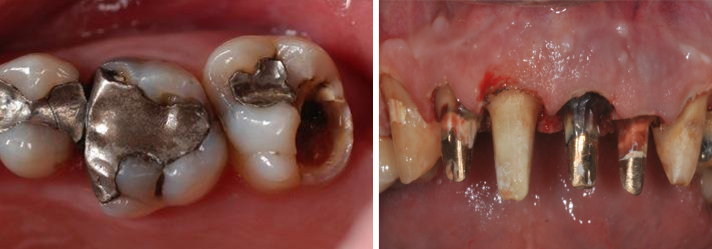
Picture: Old amalgam fillings and metal pins.
For different metals such as mercury, gold, platinum, copper, cobalt, aluminum, iron and chrome, the cytotoxic (e.g. neurological diseases), immunological (autoimmune disorders) and mutagenic (e.g. cancer) effects as well as effects on the metabolism (e.g. oxidative stress) are scientifically well documented [4–13]. Metal components can usually be detected throughout the entire body a few days after installation in the mouth.
One must always differentiate between immunological and toxicological problems. While practically every metal represents a foreign body for the immune system and exhibits allergenic potential, amalgam in particular plays a crucial role from a toxicological point of view.
Amalgam – highly toxic hazardous waste in our body.
Even today amalgam is routinely used in most dental practices. On the one hand this is because it is a material that is easily processed and holds for a long time, and on the other because it is subsidized by health insurance companies and is therefore free.
In Norway (2008) and Sweden (2009) mercury has been completely banned for some time, which of course also includes fillings. In Russia amalgam was already abolished completely at the end of the 1970s.
In contrast, the Federal Association of Dentists (BZÄK) requires amalgam as filling material.
In practice, amalgam must be disposed of after removal as a highly toxic hazardous waste – this fact alone should give pause for thought. Amalgam consists of 50% mercury (Hg), which contrary to what is often assumed is not firmly set in the filling after mixing.

Picture: Example composition of an amalgam filling: 51% mercury (Hg), 21.5 % silver (Ag), 14.8 % tin (Sn) and 12.7 % copper (Cu) (according to manufacturer’s data).
Chewing, grinding, tooth brushing and hot or cold beverages release a certain amount of mercury vapor every day. All this takes place within the microgram range; however, if one considers that even one molecule of Hg can destroy nerve cells, this cannot be underestimated. A study by Leong and Lorscheider showed that inorganic Hg quantities of 0.02 ng Hg/g led to a complete destruction of the intracellular microtubules and to a degeneration of nerve axons [14]. Hg is considered the most toxic non-radioactive element and thus exceeds all other known elements, such as lead, cadmium and arsenic, in some cases many times over [15–17]. In animal studies, after 14 days of amalgam wearing time, pathological changes could already be detected in the brain [18,19].

Picture: Smooking Tooth – www.uninformedconsent.org: Hg vapor from a 50-year-old filling – shown under fluorescent light
About 2–3µg mercury vapor is released daily per filling and that is over an average wearing time of 20 years. This therefore qualifies as low-dose, chronic poisoning. In numerous studies an approximately 2–5-fold increase in mercury in the blood and urine was observed in living amalgam carriers; investigations on deceased patients found even 2–12-fold increases in Hg levels in different body tissues. According to these studies, amalgam is the main source for mercury load in the human body [8,20–39]. Mercury can trigger any symptom and is not tolerable in the body for these reasons. The human body is extremely intelligent and stores, wherever possible, fat-soluble toxins in the metabolically inactive connective or adipose tissues. However, in athletic people or those with a low percentage of body fat, the toxins are frequently deposited in the nerve tissue or brain. In particular, babies who are nursing or even during pregnancy in the mother’s body are at risk, as Hg can penetrate the placenta. The amount of mercury in breast milk and amniotic fluid clearly correlates with the number of amalgam fillings the mother has [40–51]. Since amalgam fillings are the main source for poisoning with mercury and other heavy metals, these should be removed either in the event of chronic illness or for preventive reasons.
However, it is exactly with this removal that most mistakes are made. Usually the dentist simply drills the filling out with no safeguards because he does not know about the problem specified above (not university doctrine). Nevertheless, a very large amount of highly toxic, inorganic mercury vapor (Hg0) develops; often patients react after even a routine amalgam removal with neurological complaints, chronic fatigue, joint and muscle complaints or other recently acquired symptoms.
For this reason the removal of the fillings under absolute safeguards is indispensable.

Picture: Chlorella
Since the body stores mercury during wearing time, particularly in the brain with a half-life from several years to decades, [52-59], it makes sense to support the body after metal removal with an active heavy metal decontamination [60–64]. The brain serves here as a type of waste disposal – once in the cell, Hg vapor very quickly oxidizes through catalases to Hg2+ and can thus be covalently bound to the thiol group of proteins, which in turn changes or even blocks biological activity. In the gastrointestinal tract the Hg from amalgam is converted from microorganisms into the organic, methylated form [65–67]. Leistevuo et al. were able to prove a 2 to 3-fold increase in organic methylmercury in the saliva of amalgam carriers compared to subjects without amalgam. Fish consumption was identical in the two groups in both quantity consumed and frequency [66]. Methylated mercury in the gastrointestinal tract seems to be much more toxic than methylmercury from fish consumption, since the Hg in the fish is already bound to amino acids, whereas it is directly formed in the body and is thus is clearly more reactive [68].
A package program cannot, as can be read on the Internet, simply be implemented for heavy metal detoxification – rather these so-called detox programs can only lead to problems, because not every patient is able to simply secrete heavy metals or other toxic substances. Knowledge of the patient’s individual biochemistry as well as of possibly existing micronutrient deficits are particularly necessary for already existing health problems. In order to be able to deal with the body’s decontamination reactions, one should put themselves in the hands of experienced physicians or naturopaths.
A complete decontamination can be correctly undertaken only after the consistent removal of all interference fields in the oral cavity. This does not include only the known amalgam.
Synergy of inflammation and metals
Through the metabolism of bacteria, sulfurous protein decomposition products such as hydrogen sulfide (H2S), thioethers and mercaptans develop, which have a high bonding capacity to heavy metals [69–71]. These result in organic metal compounds of substantially increased toxicity, for example dimethylmercury [65–67] – e.g. amalgam filling on an endodontically treated tooth. Chemist Karen Wetterhan used this material in the laboratory to examine the disease-related dynamics of molecules at the cellular level. A drop of dimethylmercury fell on her latex-gloved hand and within a few hours spread through her skin to her entire body. Despite subsequent chelation therapy, she died few months later of mercury poisoning. The concentration of these poisonous molecules is many times lower through the metals in the mouth; however, it is important to know that we as dentists are exposing patients and ourselves [34] to these highly toxic substances and installing these as a source 24 hours a day, 7 days a week for the entire wearing time in the body.
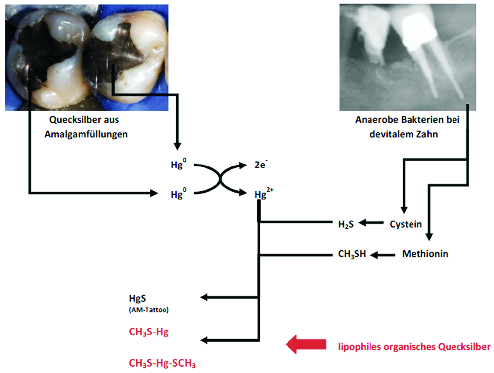
Picture: Mercury vapor from amalgam fillings can mutate in conjunction with bacterial biotoxins into highly poisonous supertoxins (dimethylmercury).
The battery effect
A battery develops when two different metals are brought into a conductive solution. The base metal ions go toward the electrochemical series in a solution and flow toward the nobler metal, electrons are released – a current flows. The saliva is the optimal electrolytic solution due to high mineral content. A classic example is a gold crown beside an amalgam filling or a gold abutment on a titanium implant. This is referred to in this case as a galvanic current or the battery effect.
These comparatively high dental mouth currents lead to the corrosion of the metals in the course of wearing time, which inevitably correlates with the problems of metal toxicity.
In addition, patients’ increasing electrical sensitivity is a result of the exponentially increasing diffusion of microwaves by WI-FI and cell phone emissions.
It must be emphasized that metals in the body act like small antennas that can disturb the sensitive action potential of the cell completely. Areas of tension develop, which sensitively disrupt the central nervous system. Inevitably one is exposed to electrical smog everywhere [72]. The standard absorption rate of electromagnetic fields can be increased 400 to 700-fold through the use of a cell phone (ringing or SMS reception) in combination with metals in the mouth [73].
Electrogalvanism and the resulting electrical sensitivity can frequently be the cause of a lack of concentration and memory loss, sleeplessness, non-specific symptoms such as a sharp pain or pressure in the chest, unexplained tachycardia, tinnitus and hearing loss, etc. [74].
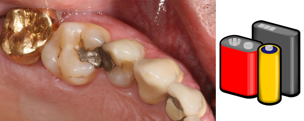
Picture: Classic scenario: Gold crown next to amalgam filling – the battery effect.
Interference fields in the oral cavity
Endodontically treated teeth
This topic is in the author’s opinion a highly significant chapter in the history of dentistry and therefore in his article “Root to Disease” it was considered in greater detail [75]. Endodontically treated teeth represent chronic-inflammatory sites that can lead to chronic problems both locally but more often in other areas of the body. More than 100 years ago Weston Price already coined the term focal infection for this purpose.
Without a blood, nerve and lymph supply the tooth is only dead organic tissue without function, which thanks to its anatomy represents the perfect cavity for pathogenic microorganisms. There are between 30 and 75,000 dentin canals per mm2. If all the dentin canals of a root were lined up, they would cover a distance of approx. one kilometer. Pathogenic bacteria exist in this widely ramified canal system of a root and form highly toxic sulfur compounds (thioethers, mercaptans) [69–71], which for their part can block vital enzymes at their active center. As nonliving organic tissue can begin to decompose over time, necrogenic material with increased toxicity (putrescine and cadaverine) can also develop.

Picture: Endodontically treated teeth on an x-ray and in real life. Dead tissue leaves traces
Inflammations at the root apex
The nonspecific immune system reacts to this infectious site with the increased production of proinflammatory cytokines (TNF-α, IL-1, INFγ). This subclinical activation of the tissue macrophages leads to chronic inflammation of the surrounding tissue and to a widening of the periodontal space and to possible cyst formation. Apart from the toxicity of sulfur-hydrogen compounds (thioethers/mercaptans), it is also common for a patient to have an allergic reaction to these substances. Root filling materials are also problematic and usually contain classic allergens such as epoxy resin, Peru balsam or rosin. Problems do not usually show up directly on site, but systemically, somewhere else in the body. Therefore, they are not always easy to diagnose.
The teeth belong to the most important subsystems within a network of self-regulating sub-ranges in the body. Teeth and their periodontium (=odonton) have a relationship with other physical structures and organs. Reinhold Voll coined the term odonton and identified the direct and close interrelations between individual odontons and the different areas of the body. Interactions and positive and negative influences in terms of a remote effect are possible in both directions:
A disturbed organ can pathologically affect the associated odonton and conversely a sick tooth or its periodontium can disturb its correlating organ. Classic interference fields next to endodontically treated teeth are displaced teeth and wisdom teeth, non-vital teeth, metal fragments and other foreign bodies, cysts and chronic inflammations in the jawbone.
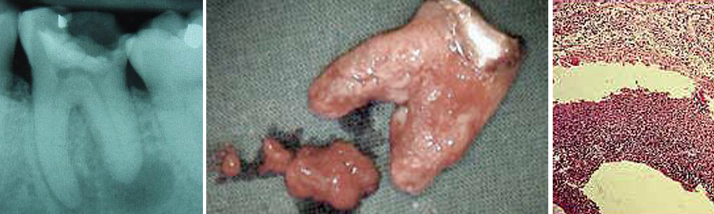
Chronic inflammations in the jawbone (residual osteitis/NICO)
Undetected by conventional x-ray, chronic inflammations in the jawbones often occur, usually resulting from old, not optimally healed tooth extraction wounds, dental germs or foreign bodies [76]. Here, similarly to endodontically treated teeth, toxic substances and inflammatory mediators (TNF-α, IL-1, RANTES) develop, which can cause various symptoms in other parts of the body – neurological (NICO) or joint problems are particularly frequent. These can be well diagnosed on suspicion when using 3D digital volume tomography (DVT).
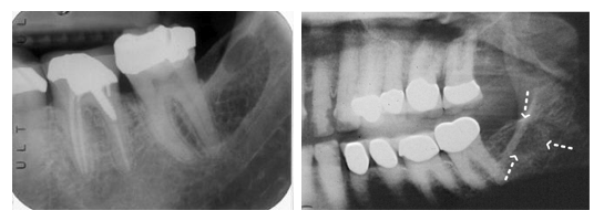
The therapy consists of the complete surgical removal of the areas altered by inflammatory activity followed by disinfection with ozone. In the author’s practice insertion of a PRGF® membrane (Platelet Rich in Growth Factors) obtained from autologous blood has proven successful. The patient’s freshly withdrawn venous blood is centrifuged for approximately 8 minutes and thus activated. After 30 minutes at body temperature, the membrane is ready to insert. PRGF® Technology is of 100% autologous origin and therefore fully biocompatible.
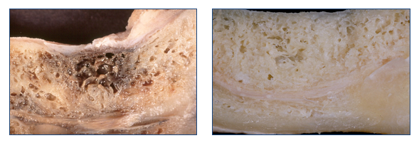
Picture: J Oral Pathol Med 1999; 28:423.

Bite problems and dysfunctions of the mandibular joint
The bite is the thermostat for the body’s structural component. The chewing process is primarily needed for our survival. In the last 100 years, in addition to iatrogenic and traumatic occlusion problems, innate and developmental abnormalities of the upper and lower jaws have become an everyday occurrence. This seems to have primarily epigenetic causes, notably nutrition plays a crucial role here. Food, mainly sugar and white flour products, seems to be directly linked to the development of malpositions of the teeth and jaws [77]. Nowadays, it has become relatively rare in industrialized nations for a child or adolescent not to have some type of orthodontic device.
Tooth and eye level are directly connected to one another: in every mammal the eyes are fixed on the horizon (labyrinthine righting reflex). Even the slightest changes in occlusion in the micrometer range lead to an imbalance in this structure. The compensation occurs through simple control mechanisms in the muscular and ligamentous apparatus, but initially through a shift of the cranium along the sutures and meninges. This mainly manifests itself in tension of the deep neck musculature; however, over a longer period it is transferred on to the shoulder and pelvic girdle. Thus, in the long-term misalignments and malpositions can develop.
The tension also leads to a reduced blood supply to the brain in certain areas as well as decreased blood flow and lymphatic drainage from the brain. Even a millimeter loss at bite level leads to a clear loss in lymphatic drainage and concomitantly to an accumulation of toxins in the brain area. Apart from the usual symptoms of craniomandibular dysfunction and myoarthropathy, a myriad of other symptoms can arise as a result, including concentration and memory loss (reduced supply of the neurotransmitter acetyl choline due to deficient blood circulation), migraine, sleeplessness, neck pain, pain in the lower back area, right up to depression [74].
The mouth could be compared to a large building site that is never finished. Every day the body must expend workers and building material in terms of nutrients and ATP in order to compensate for this building site.
24 hours a day, 7 days a week nutrients are used for the body’s ability to compensate, in other places in the body deficits or even deficiency symptoms occur. The toxic substances are increased and the body can no longer take care of the disposal – the barrel overflows. Here the key partly lies between biological dentistry and today’s common chronic illnesses. According to Straub et al., the body’s energy loss due to a chronically activated immune system is calculated at roughly 30 percent [78]. These patients are also missing a third of the energy in the morning after they get up. Small wonder then that chronic fatigue has become a widespread disease. Additionally, the body’s individual, genetic detoxification activity naturally plays an important and crucial role in this equation. This often includes inadequate nutrition, food intolerances, chronic bacterial and viral infections as well as other environmental influences (plastics, pesticides, solvents, insecticides, preservatives, etc.), to which the population is exposed daily. As such, consistent biological cleaning of the oral cavity plays an important role not only for chronically ill patients but also, if not more so, for everyone in terms of prevention.
The Alternatives
It is generally known that highly toxic mercury vapor (Hg0) develops with the removal of amalgam. Therefore, for the removal of the metals, in particular amalgam removal, maximum safeguards must be in place to protect the patient, but also above all for the dentist and his team who come into contact with the vapor daily, usually over decades.

Picture: Safeguards during professional amalgam removal. Dental dam – gold nasal hood – Clean-Up suction tip.
Not shown: oxygen – nasal probe.
In the author’s practice the following protocol has been tested:
Metal-free restorations
In biological dentistry the correct material plays a crucial role. Whether complex blood tests are done (LTT test) or whether materials are examined using bio-energy diagnostics (Autonomic Response Testing, Applied Kinesiology, Bioresonance, etc.) is up to the dentist. In an era of the hyperreactive, no longer tolerant immune system, the selection of the optimum substances should play an important role, before possibly mismatching or allergenic materials are permanently incorporated into the patient’s body. Because this could be the drop that makes the already brimming barrel overflow. It is also important to understand the entire body as an integrative system, instead of dividing it into its individual parts. In many cases it is common in the author’s practice to first shut down a primarily hyperreactive immune system through functionally medical aspects from the areas of nutritional-, environmental- and orthomolecular medicine, to remove all metals, inflammations and endodontically treated teeth and then to bring stability to this structure via long-term temporaries. Particularly with chronic progressive diseases such as MS, Parkinson’s, Alzheimer’s, cancer and ALS, this procedure is of crucial importance.
The material of choice, both for biocompatibility and aesthetics, is currently ceramic. Different types of ceramic are available to choose from: Constructions made of zirconia and more recently lithium disilicate ceramic (IPS e.max/ivoclar vivadent) have been well tested. Even the classic feldspar ceramic can be used.
The author is the Vice-President of the International Society of Metal Free Implantology (ISMI e.V.), and as such his clinical practice and all surgeries are performed free of metal.
Metal-free implants made of high-speed ceramic (zirconia) represent an important alternative to the usual titanium implants. According to Dr. Volker von Baehr (IMD Berlin), 15 to 20% of the population reacts incompatibly to titanium [79], mainly triggered by the massive use of titanium dioxide as filler or dye in medications, dietary supplements, hygiene products, cosmetics, chewing gum and toothpaste. Tissue-specific macrophages react to the titanium oxide particles primarily resulting from abrasion with phagocytosis and an increased nonspecific immune response (expression of “alarm cytokines” TNF-α, IL-1). Radar was able to show that zirconia particles of the same size in turn induce no pro-inflammatory cytokines (TNF-α) in macrophage cultures [80].
If one would also like to insert titanium for patients without risk, a titanium stimulation test should be done first in any case and a genetic predisposition to inflammation determined in the blood of patients with exaggerated immune response (high responder) to ensure that the patients will not be harmed by the implanted metal. However, the debate remains as to whether the metals work as an antenna in the mouth and thereby increase the patient’s electrical sensitivity.
Zirconia is a neutral element, translucent and biocompatible. Compared to titanium, zirconia does not possess free electrons on the surface, and therefore it cannot be in any way an interference field. Zirconia is also highly esthetic due to its white color in contrast to the grey of the titanium implants. With the help of the zirconia implant it is possible to combine biocompatibility and esthetics. Recently, zirconia implants have been available even as two-piece screw implants for all indications.
The biologically oriented dentist can also use PEEK (polyetheretherketone) screws for installation. This material has already long been used in orthopedic surgery as a replacement for intervertebral disks, since it is very similar to bone and cartilage in its elasticity. Both in Konstanz and in Tubingen, the author has used PEEK for some time for secondary frameworks, bridges or as the basis for one-piece cast prostheses. It is very flexible and can also be used from an osteopathic point of view for larger splinting, as it permits or can compensate for the independent movement of the sutures in contrast to the metal framework. Also, the chewing function benefits from this material, as its elasticity serves as a kind of shock absorber for chewing forces. This area is still new territory for dentistry; however, such innovations are needed to achieve long-term progress on the path to the optimum material; furthermore, the costs of a secondary framework made of PEEK are clearly lower than its metallic competitor.
All-ceramic materials have worked satisfactorily in the last few years not only in the areas of prosthetics and esthetics, but have for a long time also been applicable for surgical purposes. Also, the costs of the metal-free restorations were clearly reduced. In view of these facts, there is no longer any need to use base metal crowns in addition to supplies containing noble metals in the mouth or to screw gold abutments on titanium implants. Without exception the patient’s well-being should be paramount, which is possible in practically every situation with today’s existing materials and techniques.
Literature
Deutsche Zahnärzteblatt 2010, 119: 222-232
In the world of ceramic implants latest findings of research have a key role. It is important to exchange ideas with colleagues about their own experiences and patient cases from practice. Members of the International Society for Metal-free implantology e.V. (ISMI) can present their research results and case reports on ceramic implants on ismi.me, thereby supporting case exchange among colleagues. Send your text and images to a vicepresident@ismi.me. We’ll get in touch with you soon.
The International Society of Metal Free Implantology e.V. (ISMI) was established in january 2014 in Constance (Germany) with the aim of promoting metal free implant dentistry as an innovative and particularly future-oriented direction within implantology.
If you are interested in further information on an ISMI-membership click here.
Download membership application
Lesen Sie diesen Artikel in Deutsch
On 12th and 13th june 2015 the International Society of Metal Free Implantology (ISMI) held its first annual meeting in Constance. Dr. Karl Ulrich Volz, president of ISMI, welcomed 170 international participants from Spain, Belgium, Sweden, Czech Republic, United States, Switzerland, Germany, Austria, Poland and Holland. The number of participants confirmed the international claim of ISMI at its best.
In the center of the scientific program were both holistic biological aspects as well as practical experience in the daily use of ceramic implants and surgical and prosthetic concepts. The theme of the annual meeting was: ceramic implants – biological and technological basics, current standards and visions.
Impressions
The congress began on friday morning with a first pre-congress symposium on practical arrangements for the use of SDS Swiss Dental Solutions implant systems. Afterwards the participants had the possibility to join the courses „Biological Dentistry“ by Dr. Dominik Nischwitz, „Implant Surgery from A–Z“ by Prof. Dr. Frank Palm or „Injections Techniques“ by Dr. Andreas Britz. An enxclusive special on top of friday’s program were two live-operations by the companies Straumann (Prof. Dr. Palm) and SDS Swiss Dental Solutions (Dr. Volz).
Another highlight of the first day was the evening event at the „Villa Barleben“. A great atmosphere, great food and drinks and a fantastic panorama of the Alps made the evening a memorable one for all.
The second conference day was dedicated to the scientific presentations. Dr. Volz gave an overview for ceramic implants. Prof. John Ionescu spoke about „Chronic heavy metal pollution in skin and environmental diseases“. The issue was deepened by Professor Dr. Vera Stejskal with her lecture „allergy and autoimmune diseases by metal pollution“. Prof. Nendonca-Caridat from Spain explained the principles of „Biological surgery“. The second presentation block was characterizes by issues of practical use of ceramic implants (Dr. Volz and Dr. Noumbissi). In the afternoon session topics such as „Elecromagnetical fields and dental metals“ by Dr. Pascal Eppe, „Biological use of hyaluronic acid“ by Professor Dr. Marcel Wainwright, and „Lateral augmentation“ by Dr. Ernst Fuchs-Schaller were on the agenda. The conclusion of this very varied and informative program made the presentations on „17 years experience with ceramic implants“ by Dr. Ralf Lüttmann, „Marketing“ by Dr. Oliver Zernial and „Ring technology and ceramic implants“ by Dr. Bernd Giesenhagen.
The first annual meeting of ISMI was thus both in terms of the scientific program as well as with regard to the many possibilities for exchange among colleagues a very successful congress.
The company was founded in January 2014 aiming to promote the metal free implantology as an innovative and very forward-looking direction within implantology. The founders of the company are renowned implantologists from Germany and abroad. The ISMI supports its members with training opportunities and regular technical and market information.
The next annual meeting of the International Society of Metal Free Implantology will be held on 10./11. June 2016 in Berlin.
Lesen Sie diesen Artikel in Deutsch
From february 3rd to 6th 2016 the International Academy of Ceramic Implantology invites dentists and specialists to its IAOCI winter congress in Montego Bay Jamaica at the Hilton Rose Hall Resort & Spa (all inclusive).
The main topics include immediate loading of zirkonia implants, hyaluronic acid for papilla enhancements, optimizing ossteointegrations with infusions and more to come. The presentations will be held by renowned speakers, such as Dr. Mutlu Ozcan (SUI), Dr. Marcel Wainwright (GER), Dr. Ulrich Volz (GER/president of ISMI), Dr. Pascal Eppe (BEL), Dr. Dr. Vera Stejskal (SWE), Dr. Dominik Nischwitz (GER) and others.
If you have any questions concerning this congress you can visit iaoci.com or post your comment in the reply field here.

Upper Picture: © XtravaganT – Fotolia/IAOCI
The 1st annual meeting of the International Society of Metal Free Implantology (ISMI) started today at Constance Clinic. Main topic of the scientific program are both practical experience in the daily use of ceramic implants and surgical and prosthetic concepts. 200 international participants, from Spain, Belgium, Sweden, Czech Republic, USA, Switzerland, Germany, Poland and Holland are expected at Constance for the 1st annual meeting of ISMI.
On 12 and 13 June 2015, the International Society of Metal Free Implantology (ISMI) holds its first annual congress in Constance. The company was founded in January 2014 aiming to promote the metal free implantology as an innovative and very forward-looking direction within implantology. The founders of the company are renowned implantologists from Germany and abroad. The ISMI supports its members with training opportunities and regular technical and market information. The theme of the annual meeting is: ceramic implants – biological and technological fundamentals, current standards and visions.
Impressions
Speakers at the 1st ISMI meeting are international experts and also experienced users from Germany, Austria and Switzerland. In addition to the main scientific lectures on saturday, participants can expect a varied pre-congress program with seminars, live surgeries and collegial exchange of ideas as well as highlight an evening event at the highest level on friday.
Lesen Sie diesen Artikel in Deutsch
Info:
OEMUS MEDIA AG
Holbeinstraße 29
04229 Leipzig
Tel .: 0341 48474-308
Fax: 0341 48474-390
Die DVD-Reihe der OEMUS MEDIA AG zu Spezialthemen der modernen Zahnheilkunde ist durch eine neue DVD zum Thema „Biologische Zahnheilkunde“* erweitert worden. Die DVD wurde eigens für die gleichnamige dreiteilige Kursreihe mit Dr. Dominik Nischwitz/Tübingen produziert. Gezeigt werden grundlegende Therapiekonzepte und Behandlungsabläufe sowie deren Umsetzung am Patienten.
Was ist Biologische Zahnheilkunde?
Die moderne Zahnmedizin steht vor der Herausforderung, dem gestiegenen Bedürfnis der Patienten nach Gesundheit, Funktionalität und Ästhetik zu entsprechen. In diesem Sinne wird es immer wichtiger, auch ganzheitliche Aspekte in der Diagnostik und Therapie zu berücksichtigen und aktiv zu nutzen. Neben der klassisch handwerklich geprägten Zahnmedizin werden bei der Biologischen Zahnheilkunde zusätzlich die naturwissenschaftlichen Grundregeln in ihrer Komplexität und Wechselwirkung im Hinblick auf den menschlichen Organismus berücksichtigt. Ganz unter dem Motto „Der Mund als Spiegel für die Gesundheit“ werden so die Grundprinzipien der Chemie, Physik und Biochemie genauer unter die Lupe genommen und ein Zusammenhang zwischen den heute üblichen chronischsystemischen Erkrankungen und Erkrankungen der Mundhöhle hergestellt.
Hinweis: Für die Teilnehmer des Spezialistenkurses „Biologische Zahnheilkunde“ ist die DVD in der Kursgebühr inkludiert.
Spieldauer 80 Minuten
* Die Ware ist vom Umtausch ausgeschlossen!
Die DVD ist im Shop der OEMUS MEDIA AG erhältlich.
Flyer zur Kursreihe als PDF.
Bestellen Sie online oder per:
E-Mail: grasse@oemus-media.de
Tel: +49 341-48474-201
Fax: +49 341-48474-290
The 1st annual meeting of the International Society of Metal Free Implantology (ISMI) will be held on 12 and 13 June 2015 at Constance Clinic. Main topic of the scientific program are both practical experience in the daily use of ceramic implants and surgical and prosthetic concepts.
On 12 and 13 June 2015, the International Society of Metal Free Implantology (ISMI) will hold its first annual congress in Constance. The company was founded in January 2014 aiming to promote the metal free implantology as an innovative and very forward-looking direction within implantology. The founders of the company are renowned implantologists from Germany and abroad. The ISMI supports its members with training opportunities and regular technical and market information. The theme of the annual meeting is: ceramic implants – biological and technological fundamentals, current standards and visions.
Speakers at the 1st ISMI meeting are international experts and also experienced users from Germany, Austria and Switzerland. In addition to the main scientific lectures on saturday, participants can expect a varied pre-congress program with seminars, live surgeries and collegial exchange of ideas as well as highlight an evening event at the highest level on friday.
Info:
OEMUS MEDIA AG
Holbeinstraße 29
04229 Leipzig
Tel .: 0341 48474-308
Fax: 0341 48474-390
ismi.me/events/1st-annual-meeting-ismi/
I. Introduction
Zirconium Dioxide (ZrO2) – or “Zirconia” as it is more commonly known, was discovered in 1789 by the German chemist M. H. Klaproth. However, it was only introduced into dentistry a few decades ago: a product of the increasing desire for highly esthetic restorations. Zirconia became an attractive alterative material in dentistry because of its high esthetic potential and comparable strength to traditional metals. In the field of implant dentistry, Titanium has been the mainstay in implant manufacturing; however, Zirconia became a viable option because it possesses superior properties, including a higher tensile strength, compressive strength, and modulus of elasticity when compared to either Titanium alloy or commercially pure Titanium (Table 1). The Zirconia used in dentistry today is not merely the Zirconium dioxide discovered in the 18th century; the commercial grade Zirconia has several modifications that enhance its properties. In its pure phase, Zirconium dioxide has a low shear strength and is very brittle, essentially making it useless as a dental material. The addition of small amounts of Aluminum Oxide and Yttrium Oxide increase the modulus of elasticity and help to stabilize the material. This combination of oxides is mixed in the powder state and placed in a sintering oven to produce a monocline crystal structure, with equally-spaced, non-overlapping particles (Figure 1). Although the monocline crystal is a strong material, cracks can propagate easily in the structure which makes it less desirable to use as a long-term implanted prostheses. In order to eliminate this issue, today’s Zirconia is also put through a process known as “HIP” (hot isostatic pressing). The high pressure under which the monocline Zirconia is placed during HIP processing causes condensation of the particles and results in a tetragonal crystalline structure, where the particles appear to overlap (Figure 2). The significance of this innovation is that it imparts the ability to stop crack propagation. When the surface of HIP-processed Zirconia is prepared, any microcracks that may result are quickly stabilized as tetragonal particles expand to the monocline structure and fill the void. The self-repairing property is also known as the “airbag effect”. The additional stability gained by the HIP process has enabled Zirconia to be used for multiple medical prosthetic devices including auditory, finger, and hip prostheses.
Zirconia dental implants have been available on the commercial market since 2004. The current major players in the United States are Bredent white SKY, ZSystems Z-Look3, and Oral Iceberg CeraRoot. Zirconia has proven its utility in dental implants through a series of animal and human clinical studies wherein it has been shown to successfully osseointegrate into bone and be highly biocompatible.


II. Indications and Contraindications for Zirconia Implants
A. Indications:
1. All esthetic zone cases, especially in those with scalloped, thin biotype gingival architecture and in critical gingival papilla build-up cases
2. Patients with metal allergies and chronic diseases resulting from them
3. As an alternative to Titanium dental implants in any intraoral location
B. Contraindications:
1. Patients that exhibit a lack of compliance to post-operative instructions.
2. A lack of operator clinical and technical knowledge about implant surgery and prosthetic restorations
3. Any other general contraindications to implant rehabilitation with one or twopiece titanium implants, such as bruxism.
III. Bone Relationship
A. One-Piece Implant Concept
The one-piece component implant was conceived in an attempt to copy nature. Imagine an implant restoration without micro gaps, internal screws, or inner gaps. The one-piece implant allows axial forces to be applied into a solid structure without attachments, made entirely of one material with no physical interruption and excellent flexural strength. One of the major advantages of the “HIP” processed Zirconia is its ability to be prepared intraorally, as ceramics do not conduct heat like metal or natural tooth structure. Preparation of the abutment can occur immediately after insertion or after osseointegration, and allows what is essentially a custom abutment to be prepared. Despite these apparent advantages, there has been a well-documented history of unsuccessful one-piece titanium implant systems, which has hindered the ability of the Zirconia onepiece implant gain acceptance into the implant community. These Titanium one-piece systems were often used to also provide immediate loading, which has not been shown to provide predictable success. Unlike the Titanium one-piece implant, the goal of the Zirconia one-piece is not to provide immediate loading, but to provide immediate esthetics. One should also consider the differences in the cost of manufacturing and the environmental implications for one and two-piece implant systems. Certainly the engineering of the many intricate components of the two-piece implant system is fascinating, but it creates exponentially more leftover material and pollution to process the much greater number of parts. The implant industry is an ever-expanding, multi-million dollar enterprise that may see little benefit from reducing the number of parts involved in implant systems, as this may in turn reduce profit. Despite this, the need for more efficient and environmentally friendly industrial operations is critical and the push towards a more economical solution will continue. In the world we’re living in today, my opinion is that less is more and that a one-piece implant is an all-around better solution, like the one-piece tooth we all are born with.
B. Importance of Proper Planning
Whereas a two-piece implant system can compensate for implant body positioning by utilization of custom-made angled abutments, onepiece implants have an inherent limited compensation ability. Proper implant positioning at the time of insertion is critical to the success of the restoration and esthetics of the final product. The abutment in a one-piece system can account for only around 20º of correction through preparation of the coronal aspect. In order to properly determine the ideal implant location, wax-ups and digital prototypes should be utilized when possible. When proper implant placement is achieved, the abutment will be in such a position that forces transmitted along the long axis will be favorable, and the unfavorable loading will be minimized. Such a relationship can then translate into a good long-term marginal bone level stability and a healthy, durable restoration.
C. Tulip-Shaped Abutment Neck
The tulip-shaped neck of the abutment is analogous to the cervical shoulder area of the implant. This area marks the transition between the implant and the abutment. It allows the implant to be inserted at a variable depth to establish the proper emergence profile with optimal gingival contour, and also enables correction of axial divergence by up to 20%. The design and material of the implant allows vertical placement in bone to vary by up to 1.5 mm. Since the Zirconia is white, there is little esthetic risk from not sinking the implant deep enough. If the crestal bone architecture is flat, the implant shoulder does not have to be countersunk, although the biologic width must be observed. In this situation, the implant should be inserted in the bone up to the coronal end of the threading. Use of the countersink is not necessary to achieve this position. For esthetic reasons such as thickness of mucosa, need for vertical adjustment of the preparation border, or with uneven crestal bone architecture it is frequently necessary to countersink the implant up to the transition of the implant tulip to a maximum of 1.5 mm. When attempting to place immediate implants in the esthetic zone, the shoulder or tulip insertion should extend to cover the edge of the extraction socket to achieve greater stability and the same results as tapered implants. After 5 years of clinical use and studies, the current recommendation is to try to avoid over-insertion of the shoulder when not needed in non-esthetic areas, as it may lead to a greater degree of bone loss over time.
D. Angled Abutments
The anatomical shape of the alveolar bone dictates the ideal position of the implant body, and maintenance of the buccal cortex is essential for the long-term success of the implant restoration. When placing implants in the anterior region, the operator often has a tendency to base the implant angulation off of the future restoration, which can consequently lead to buccal cortex violation. With the implant body at the correct angulation, the restorative components may not be properly angled for a good esthetic result; often the abutment protrudes buccally leaving little room for fabrication of a naturalappearing crown. Two-piece implant systems may use angled abutments to compensate for this discrepancy. In one-piece Zirconia implants, the issue is easily addressed by preparation of the abutment aspect to the desired angle, up to a maximum of 20 degrees. This is possible because the wide implant shoulder in combination with the large abutment allows an even force distribution, which minimizes bone loss and increases longevity of the restoration.
IV . Soft Tissue Relationship
A. Zirconia surface
The Zirconia surface of the implant is biocompatible with the oral soft tissues. As a ceramic, Zirconia inhibits formation of plaque and promotes a healthy soft tissue attachment. There has been no evidence of any inflammatory reaction or irritation to the gingiva from the Zirconia surface.
B. Scalloping the implant shoulder
The implant shoulder may be adjusted to better follow the scalloping of the gingiva to obtain the most esthetically pleasing results in the anterior region.
C. No micro gaps
Eliminating the micro gap between the implant body and abutment eliminates the possibility of bacterial attachment and inflammation. Without a micro gap, there is less long-term soft tissue irritation.
D. Gingival papillae growth
The gingival soft tissues have been found to have an affinity for the Zirconia surface, which leads to excellent esthetics. Not only can the Zirconia preserve the existing gingival papilla height, but it has even been observed to induce gingival growth. For papilla build-up cases, Zirconia therefore has a distinct advantage over traditional Titanium implants. The best results have been shown in cases with a thick and flat gingival biotype as well as a good emergence profile without violation of the biologic width.
E. Surgical Considerations
For the best esthetic results, one should start the process of contouring the soft tissues at the time of tooth extraction during immediate placement, and when the provisional is first made during the traditional protocol. When planning an immediate placement case, a conservative, atraumatic extraction will aid tremendously in maintaining the best gingival architecture. The provisional should have a smooth and well-contoured finish line to facilitate the best gingival health. Often the tissues will be inflamed at the time of surgery, especially with immediate implant placement, because of pre-existing infection in the tooth. It is quite common, therefore, to have what appears to be recession of the tissues during the healing process. As the Zirconia surface is biocompatible and does not trap plaque, the tissue inflammation will subside in 1-2 weeks after placement. Flapless surgery is a good alternative to help with soft tissue maintenance. Flapless procedures are usually done with the help of a tissue punch, and require good case planning for proper implant placement. There is a range of approximately 1.5 to 2 mm of acceptable variation in the vertical insertion depth of the implant shoulder. The soft tissue biotype, height of the bony crest, and soft tissue thickness must all be taken into account to obtain the best implant position and subsequently the correct emergence profile.
V. Intraoral Adjustments
A. Implant selection
As with any treatment planning for any type of dental implants, several factors must be taken into consideration when planning for one-piece Zirconia implants. The total number of implants, diameter, length, and position should all be based on the available space, quantity, and quality of bone. The minimum height required for one-piece Zirconia is thought to be 7 mm. Bone grafting procedures should be undertaken when necessary to achieve this minimum height. If the crestal bone architecture is flat, the implant does not need to be countersunk; however, if the soft tissue esthetics dictate that the implant must be countersunk, it may be placed up to 1.5 mm deeper than the last thread. All Zirconia one-piece implants should be surrounded by at least 1.5 mm of bone, with 3 mm of bone between two implants. The implant diameter should be based on the tooth being replaced, anticipated occlusal forces, and the available space between the roots of neighboring teeth. The minimum distance of the implant shoulder to the adjacent teeth is 0.5 mm, measured from the greatest curvature of the neighboring teeth, keeping in mind that the implant shoulder can be adjusted up to 1 mm when necessary.
B. Abutment preparation
After insertion of the one-piece implant, it may be necessary to prepare the abutment to meet the anatomical demands of the site. Ideally all biting forces should be directed along the long axis of the implant, but the abutment aspect of the implant may be prepared to compensate for angulations up to 20º. When available, wax-ups should be used to aid in treatment planning. When adjusting the abutment immediately after implant placement, red-striped ultra fine grain (46 μm) diamond burs should be used to a maximum bur speed of 160,000 rpm. A minimum of 50 ml/min of irrigation should be utilized during the procedure, and excessive forces should be minimized on the newly placed implant. The abutment should only be prepared enough to allow for adaptation of the provisional restoration, as more definitive adjustments will be made following soft tissue healing. If the shoulder needs to be lowered in the mesial or distal aspects of the site, this should be completed prior to closure of the soft tissues. As the provisional restoration will need to be out of occlusion, the abutment should be a minimum of 1.5 mm below the plane of occlusion, but no less than 3 mm in height. After the healing phase and implant osseointegration, the definitive preparation of the implant shoulder can be completed. At this time, one should ensure proper scalloping of the shoulder to match the soft tissue contours and the abutment should be properly rounded prior to the final impressio.
C. Inserting the implant and increase the bone to implant contact
One of the key factors in dental implantology is a good primary stability. What we considered in our learning curve is that we increased the bone to implant contact by condensing the spongy bone. Depending on the bone we did drill with the final drill only through the corticalis and not the spongiosa anymore. By inserting the implant with a higher torque (up to 45-50 Ncm) we compressed the spongy bone with the implant and increased in the spongy bone a higher bone to implant contact. Please make sure using this technique only for the spongiosa.
VI. Guidelines for an ideal emergence profile
A. Gingival Biotype
The thick and flat gingival biotype offers the best overall esthetic results, including the best coverage of the margin and papillae preservation. The thin and scalloped biotype makes it more challenging to adjust and maintain the best cervical margin; however, using Zirconia implants eliminates the problem of the grey gingival shadow associated with Titanium implants. If recession occurs and exposes the crown margin, although less esthetically pleasing, it will not be as undesirable as an exposed Titanium surface.
B. The Zirconia Implant surface characteristics
Zirconia has been shown through a number of clinical studies to have great tissue biocompatibility and long-term stability. When in contact with tissue fluids, the implant surface carries a neutral polarity which disables bacterial aggregation. This, in combination with the lack of a micro gap, makes the one-piece Zirconia implant a great tool for management of the soft tissues. These characteristics allow for excellent gingival health and even spontaneous growth of soft tissues, which is an advantage for the long-term esthetics of dental implants.
C. Bone and soft tissue level
Just as with any dental implant, the best esthetics will be achieved when the implant has good bony support on all four walls. Clearly this is best accomplished with an atraumatic extraction and ideal placement of the implant, but when this is not possible, bone grafting may be necessary. If a significant amount of marginal bone is lost during extraction or there is a vertical discrepancy in ridge height compared to adjacent teeth, an implant restoration will require a longer crown to compensate. This situation should be avoided in the esthetic zone, particularly in patients with a high smile line. If a one-wall or small volume defect is present and the patient is planned for immediate implant placement, bone grafting material may be used, which is very wellaccepted by Zirconia implants. For larger defects where a significant volume of bone is missing, a two-stage procedure should be undertaken and implant placement delayed until completion of grafting.
D. Implant positioning
The ideal emergence profile of an implant will be created by placing the implant in its ideal position. Selecting the proper implant diameter is a vital part of this process. By using a small diameter implant to support a large restoration, we create an unnatural and unesthetic emergence profile. Implant diameters must be properly matched with the size of the interdental space to be restored. Implants must also be placed in their ideal vertical position to achieve proper emergence. For the one-piece Zirconia implants, there is a range of 1.5 mm in vertical positioning for which ideal esthetics can be maintained. Necessity of countersinking is situation-specific and depends on operator preference, but in general is necessary when the crestal bone is thin or irregular or soft tissues are very thin. Implants can be countersunk so that the implant neck is partially embedded in crestal bone and the shoulder remains subgingival; they may be countersunk from the last coronal thread up to half the height of the implant neck.
E. Implant Preparation
Customizing the abutment portion of the one-piece implant is a technically simple procedure that is generally very quick. After implant placement, gross discrepancies can be adjusted if necessary, bearing in mind that tissues will likely undergo some degree or recession. One must also take into account that adjustments made will be irreversible, and it is always preferable to defer adjustment until after healing has occurred. Ideally, implants are prepared after osseointegration and tissue remodeling has been completed. The implant shoulder should be scalloped to match the gingival contour of the tissues and allow for subgingival placement of the crown shoulder. The recommended shoulder design is a chamfer, which can be easily created with a Torpedo ISO 016. The maximum speed of rotary instruments used on Zirconia implants is 160,000 rpm with copious irrigation. Other important adjustments include angulation of the abutment portion to match adjacent teeth and creating a common path of insertion for multi-unit prostheses. Narrow neck implants, which are designed without a clear marginal line, may require less or even no intraoral adjustments. When necessary, they can be prepared with the flame ISO 012 for a knife-edge type shoulder design.
F. Provisionals
Most Zirconia implants are temporarily restored after placement with provisional restorations. Provisionals should be well-adapted and polished so as not to irritate the tissues and hinder the healing process. Since the implant shoulder will be slightly subgingival, so must the provisional. It should have good circumferential contact with the shoulder and be wide enough to allow the tissues to heal with the proper contour for emergence and to maintain papillae architecture. The operator should consider changing or rebasing the provisional restoration after approximately 3 weeks of healing to aid in soft tissue management. After this time, the tissues wi l l be approaching their final conformation, and additional contouring of the provisional wi l l allow for any necessary adjustments to soft tissue shape.
VII. Common Mistakes
A. Incorrect Implant Positioning
One-piece implants demand accuracy in placement due to our limited ability to compensate for mistakes as compared to twopiece implant systems. It is important to plan properly and use advanced planning techniques such as cone-beam CT, digitally guided implant placement, and surgical guides whenever possible. Improper placement can lead to nonrestorable implants, apical exposure, proximity to adjacent roots, or unfavorable forces on the restored implant.
B. Premature Loading
The design of the one-piece implant dictates that the restorative abutment be present and exposed to the oral cavity from the time of placement. This leaves the implant vulnerable to premature forces during the healing period which can potentially compromise osseointegration. Chewing, cheek pressure, and tongue pressure can all cause implant micro-movements that may lead to failure of the integration process. In order to adequately protect the implant, there are a variety of provisional restorations that can be employed, including an Essix appliance, eggshell temporary, reworked denture, Maryland bridge, posterior adhesive bridge, or thermoplastic clasp denture. The success of the implant is highly dependent on adequate protection during the integration period and a proper protective device should be fabricated within the first 24 hours. When possible, a premade provisional that can be adjusted at the time of implant placement is preferable. The device should provide 1 to 1.5 mm of free space circumferentially around the abutment and be out of occlusion during all functional and parafunctional movements.
C. Improper Abutment Preparation
Poor abutment preparation may lead discrepancies in spacing or angulation. If the implant is prepared in such a way that one side of the abutment is trimmed much more than the other, the resulting crown may not be balanced over the implant and deleterious forces may be transmitted.
D. Incorrect Implant Width
As with traditional dental implants, the mesiodistal width of the site for implant placement should provide at least 1 mm of bone between the implant and adjacent teeth. In order for the one-piece implants to be placed, including the wider shoulder area, the important area to measure is between the height of curvature of the adjacent teeth. There should be a minimum of 0.5 mm on either side of the implant to allow placement. With less than 0.5 mm of space, esthetics will be compromised and the patient may have difficulty appropriately cleaning the area. In addition, ingrowth of papillae may be truncated, which would also negatively impact the esthetic outcome.
E. Lack of Consideration for Soft Tissue Biotype
When treatment planning for implants, the soft tissue biotype must be taken into consideration. Failure to address this issue can lead to less than ideal esthetic outcomes that may have been easily avoided. For one-piece Zirconia implants, one important consideration is that implants should be countersunk in those with a thin and scalloped gingival biotype. The implant shoulder should be inserted into the bone as deep as possible to attain a suitable cervical emergence profile. By misjudging or neglecting to consider the gingival biotype, one may end up excessively grinding the implant shoulder to attempt to place the finish line in a subgingival location. Often the result is an unesthetic supragingival finish line and poor papillae ingrowth.
VIII. One–piece implants Vs. Two-piece implants with Zirconia Abutments
A. Clinical benefits of One-piece Zirconia Systems
1. Having a single stage procedure
2. Decreased chair time – preparing the abutment is similar to preparing a traditional crown, and intuitive for the experienced operator.
3. Less complex armamentatium, fewer parts required for restorative procedures
4. Elimination of lab time for abutment fabr i cat i on; No need for healing abutments, screws, analogs, or transfer copings.
5. No internal screws, no internal gaps, no micro gaps, fewer locations for hardware failures.
6. Excellent soft tissue integration.
7. Less consequences from gingival recession. As minor recession may occur over time when chewing forces are extreme, and exposure of the white implant surface will be less noticeable than Titanium.
8. No gray gingival showthrough. Zirconia implants will never cause the unesthetic gray gingival tint which attracts the eye and makes Titanium implant restorations in the esthetic zone very challenging.
9. Flexural Strength. Zirconia is bone friendly and has good flexural strength for supporting implant prostheses.
10. Improved gingival health. Due to the lack of micro gaps and the neutral polarity of the implant surface, there is much less tendency to have an erythematous, inflamed implant neck as is often seen after removal of the healing abutment with Titanium implants.
11. Force distribution. The one-piece design allows force to be transmitted equally throughout the structure.
12. No metal parts. Zirconia is the only material outside of Titanium being used for dental implants. This is advantageous for those with metal allergies or with an interest in holistic dentistry.
B. Clinical Disadvantages of One-piece Zirconia Systems
1. The implant must be protected during healing.
2. Less ability to compensate for incorrect implant angulation.
3. Necessity for good patient compliance. Not every patient may be able to be compliant enough to allow for the necessary healing phase without force being applied to the implant.
4. The healing process may last from 3 to 6 months, depending on bone quality.